Pediatric Vision Screeners (developed at
the Wilmer Eye Institute)
Pediatric Vision Screeners developed by us check mainly for risk factors for amblyopia. Amblyopia, commonly known as “lazy eye,” is poor transmission of the visual image to the brain. It is estimated to afflict up to 3.6% of children, and is among the three leading causes of visual impairment in adults. It can be successfully treated, but only early in childhood, especially during infancy. Delayed treatment, on the other hand, risks lifelong visual impairment. Early detection of the primary causes of amblyopia – strabismus (misalignment of the eyes) and defocus – is therefore extremely important, yet often difficult in young children and infants, especially by untrained individuals. A new generation of computerized electro-optical instruments that test for the presence of amblyopia’s major risk factors heralds the potential for screening that is both simpler to administer and more reliable than traditional sensory testing. However, this new generation of screening technology in currently available implementations has serious limitations, especially in terms of sensitivity and specificity. Some vision screening devices can assess fixation of the eyes (eye alignment) only indirectly via highly insensitive analysis of the position of the corneal light reflexes, involving a calibration step with each eye looking in a known direction.
Our laboratory has been developing a novel technology for detecting accurate eye alignment directly, by exploiting the birefringence (property that changes the polarization of light) of the uniquely arranged nerve fibers (Henle fibers) surrounding the fovea (the small area of the retina, responsible for sharp central vision). Retinal birefringence scanning (RBS) is a technique that uses the changes in the polarization of light returning from the eye to detect the projection into space of the array of Henle fibers surrounding the fovea. In RBS, polarized near-infrared light is directed onto the retina in a circular scan, with a fixation point in the center, and the polarization-related changes in light retro-reflected from the ocular fundus are analyzed by means of differential polarization detection. Due to the radially symmetric arrangement of the birefringent Henle fibers, a characteristic frequency appears in the obtained periodic signal when the scan is centered on the fovea, indicating central fixation. By analyzing frequencies in the RBS signal from each eye, the goodness of eye alignment, and thus strabismus, can be measured. In preliminary studies and in an early prototype, RBS has demonstrated reliable and non-invasive detection of foveal fixation, as well as detection of eye misalignment.
Our goal is to create a new instrument that will make it possible to screen for the risk factors for amblyopia both earlier and more reliably than has previously been possible. This instrument will, we believe, enable earlier treatment and lead to an improved clinical outcome for these children. It has the potential to prevent millions of children from developing lifelong disability from a readily treatable cause of impaired vision.
The 2002 PVS
prototype:
|
|
|
|
RBS / central fixation detection |
Focus detection |
The design process
The design process starts with defining the exact purpose and function of the device and building the first laboratory prototypes as research instruments. These instruments are then tested (validated) in the lab and on a small number of patients, after obtaining institutional approval (IRB). So far we have successfully developed and tested two such prototypes – PVS1 and PVS2.
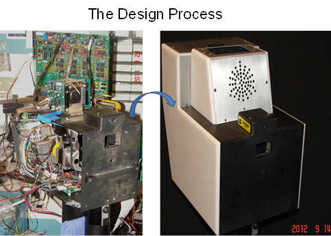
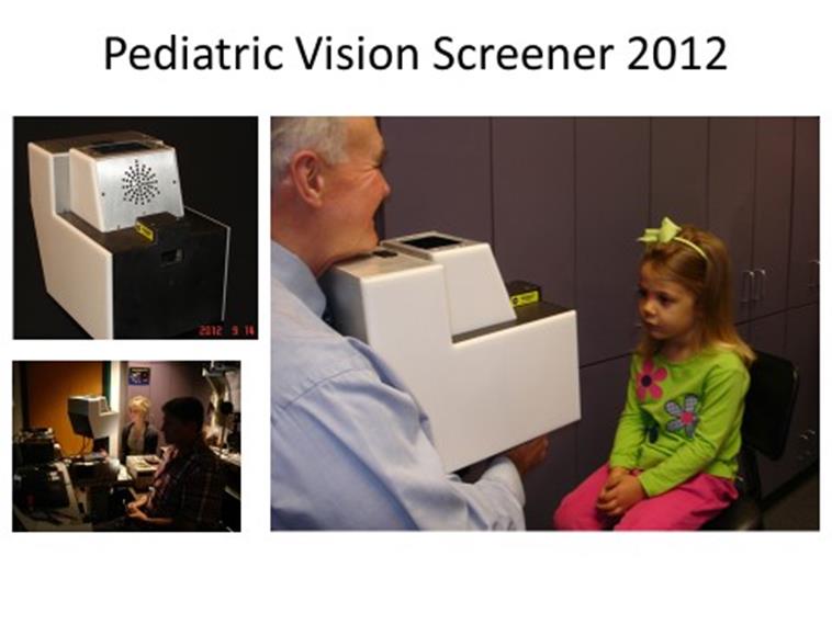
The 2012 prototype of the PVS. Shown are parts of the optics, electronics and, the optomechanics and the enclosure. The device has institutional safety clearance by the IRB and Clinical Engineering. It is being tested rigorously on young amblyopic anc healthy control subjects.
Operating the device
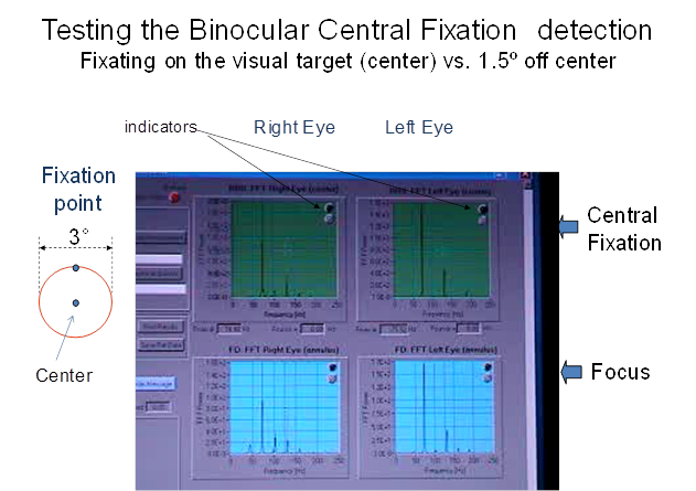
The panel below show the graphical user interface (GUI) of the PVS 2012. If during the exam central fixation or defocus is detected for any eye, the respective virtual LED indicator changes from green to red. The custom software for the GUI and real time operation has been developed in our lab. Results are archived for further statistical and optimization analysis.
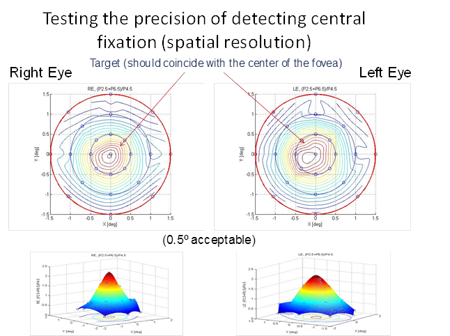
Device verification
The
test subject was asked to at different points in the close vicinity of the
fixation target. For each point, the RBS signal was measured and plotted.
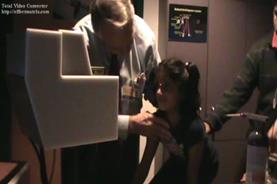
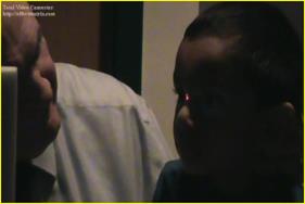
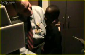
PVS 2012 - Device testing on
pediatric subjects
|
|
|
|
|
Dr. David Guyton conducting the first tests of the device |
||
|
|
|
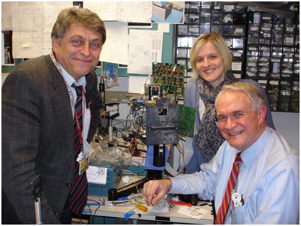
The main team: Drs. David Guyton, Boris Gramatikov and Kristi Irsch |
References
- Nassif D.S., Piskun N.V., Gramatikov B.I., Guyton D.L., Hunter
D.G. Pediatric Vision Screener 2:
pilot study in adults. Journal
of Biomedical Optics, 2004 Nov;9(6):1369-1374,
http://www.ncbi.nlm.nih.gov/pubmed/15568960
- Hunter D.G., Nassif
D.S., Piskun N.V., Winsor R., Gramatikov B.I.,
Guyton D.L. Pediatric Vision
Screener 1: instrument design and operation. Journal of Biomedical Optics,
2004 Nov;9(6):1363-1368.
http://biomedicaloptics.spiedigitallibrary.org/article.aspx?articleid=1101874
- Nusz K.J., Congdon N.G., Tang Ho, Gramatikov B.I., Friedman D.S.,
Guyton D.L., Hunter D.G. Rapid,
objective detection of cataract-induced blur using a bull's eye
photodetector. J Cataract Refract Surg. 2005
Apr;31(4):763-70,
http://www.ncbi.nlm.nih.gov/pubmed/15899454
http://www.sciencedirect.com/science/article/pii/S0886335004009484
4.
Gramatikov B.I., Zalloum
O.H.Y., Wu Y.K., Hunter D.G., Guyton D.L. Birefringence-based eye
fixation monitor with no moving
parts. Journal of Biomedical Optics, 2006, 11(3), May-June, pp. 034025-1 -
034025-11,
http://www.ncbi.nlm.nih.gov/pubmed/16822074
5.
Irsch,
K., Gramatikov, B., Wu, Y.-K., Guyton, D.
Modeling and minimizing interference from corneal birefringence
in retinal birefringence scanning for foveal fixation detection. Biomed
Opt Express. 2011 Jul 1;2(7):1955-68.
http://www.opticsinfobase.org/boe; http://www.ncbi.nlm.nih.gov/pubmed/21750772
6. Gramatikov,B., Irsch,K., Müllenbroich,M., Frindt,N., Qu,Y., Gutmark,R.
Wu,Y.-K., Guyton, D. A Device for Continuous
Monitoring of True Central Fixation Based on Foveal Birefringence. Annals of Biomedical Engineering, Vol.
41, Issue 9 (1 Sept 2013), pp. 1968-1978.
Also Epub ahead of print: May 4, 2013. DOI:
10.1007/s10439-013-0818-2. PMID: 23645511.
http://link.springer.com/article/10.1007%2Fs10439-013-0818-2
7.
Irsch,K., Gramatikov,B.I., Wu, Y. K., Guyton,D.
L. A new pediatric vision screener
employing polarization-modulated, retinal-birefringence-scanning-based strabismus
detection and bull’s eye focus detection with an improved target system: Opto-mechanical design and operation. Journal
of Biomedical Optics, 2014, Jun 1;19(6):67004. doi:
10.1117/1.JBO.19.6.067004.
http://biomedicaloptics.spiedigitallibrary.org/article.aspx?articleid=1881172
- Irsch,
K, Gramatikov, B.I., Wu, Y-K, Guyton, D.L. Improved eye-fixation
detection using polarization-modulated retinal birefringence scanning,
immune to corneal birefringence. Optics Express,
22(7):7972-7988, published on 18 March 2014,
DOI:10.1364/OE.22.007972, http://www.opticsinfobase.org/oe/abstract.cfm?uri=oe-22-7-7972
http://www.opticsinfobase.org/oe/fulltext.cfm?uri=oe-22-7-7972&id=282349